• 94150 94782

A-Scan

At Deep Netralaya, we offer the latest in diagnostic technology to ensure the highest level of accuracy in assessing and treating eye conditions. One such advanced diagnostic tool we use is the A-Scan (also known as an A-Scan Ultrasound), which is an essential test for evaluating the health of your eyes and determining the proper treatment

Why Choose Deep Netralaya for Your A-Scan?

  • State-of-the-art Technology: We use the latest, high-quality A-scan machines to ensure the most accurate readings.
  •  
  • Expert Care: Our skilled team of ophthalmologists and technicians are trained in using this technology to provide you with the best care possible.
  •  
  • Patient Comfort: We prioritize your comfort and make sure the procedure is done smoothly and efficiently, with no discomfort or downtime.
B-Scan

At Deep Netralaya, we believe in using the latest technology to provide our patients with the most accurate diagnoses and effective treatments. One of the essential diagnostic tools we use is the B-Scan Ultrasound, a non-invasive imaging technique that plays a crucial role in evaluating the internal structures of the eye, especially when other methods are not sufficient.

Why Choose Deep Netralaya for Your B-Scan?

  • Advanced Technology: We use cutting-edge B-scan machines to provide high-resolution images for accurate diagnosis and effective treatment.
  •  
  • Expert Ophthalmologists: Our experienced specialists are skilled in interpreting B-scan images and tailoring treatment plans based on the findings.
  •  
  • Comprehensive Care: We provide a full range of eye care services, ensuring that every aspect of your eye health is carefully monitored and treated.
  •  

NCT (Non contact tonometry) Pachymetry

At Deep Netralaya, we utilize the latest diagnostic technologies to provide our patients with the most accurate and comfortable eye care. One such advanced tool is Non-Contact Tonometry (NCT), a vital procedure for measuring the intraocular pressure (IOP)—a key factor in diagnosing and managing glaucoma.

Why Choose Deep Netralaya for Your Pachymetry?

  • Precision Technology: We use state-of-the-art pachymetry devices to provide precise and reliable measurements.
  • Comprehensive Care: Our ophthalmologists utilize pachymetry results to create individualized treatment plans tailored to your specific eye health needs.
  • Expert Team: Our specialists are experienced in interpreting pachymetry results and offering the best course of action to ensure your eye health is carefully monitored and managed.

Pachymetry

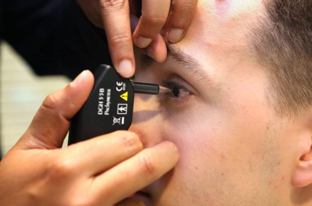
Pachymetry, particularly in the context of diagnostic practices at specialized eye care centers like Deep Netralaya (a name that could refer to a particular eye hospital), is crucial for a range of eye health assessments. Eye centers typically use pachymetry to measure corneal thickness to aid in the diagnosis and management of several ocular conditions

Use of Advanced Pachymetry Techniques

  • Ultrasound Pachymetry: This is an accurate, widely-used method where an ultrasound probe is placed on the cornea to measure its thickness.
  • Optical Pachymetry: Non-contact methods that use light to measure thickness without touching the cornea.
  • Ocular Coherence Tomography (OCT): Some centers might use OCT to obtain high-resolution images of the cornea and measure its thickness more precisely.

AT(Applanation tonometry)

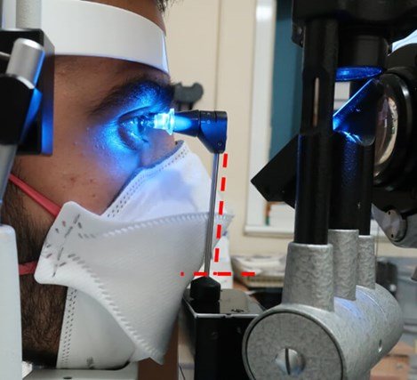
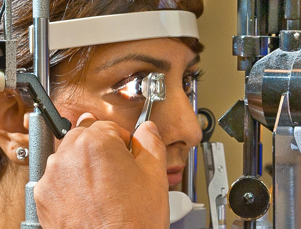
Applanation Tonometry (AT) is a method used to measure intraocular pressure (IOP), which is the fluid pressure inside the eye. This measurement is important for diagnosing and managing conditions such as glaucoma, where elevated IOP can lead to optic nerve damage and potential vision loss.

Procedure

  • The patient is typically seated in front of a slit lamp.
  • Topical anesthesia (eye drops) is applied to numb the surface of the eye to minimize discomfort.
  • A small amount of fluorescein dye is instilled into the eye to make the cornea visible under the slit-lamp light.
  • The tonometer probe touches the cornea lightly, and the device measures the force required to flatten the cornea. The IOP is then calculated.

Gonioscopy

Gonioscopy is a diagnostic procedure used to examine the anterior chamber angle of the eye, which is where the cornea and iris meet. This angle is important in understanding how fluid drains from the eye. Gonioscopy helps in assessing conditions that affect the drainage system of the eye, such as glaucoma

Purpose:

  • Glaucoma Diagnosis: The primary use of gonioscopy is in diagnosing types of glaucoma, especially angle-closure glaucoma, where the angle between the iris and the cornea becomes blocked, leading to a sudden rise in intraocular pressure.
  • Identifying angle-closure: It helps distinguish between open-angle glaucoma and angle-closure glaucoma, which requires different treatments.
  • Pre-surgical Evaluation: Gonioscopy can be useful for evaluating the anatomy of the anterior chamber angle before certain types of surgery, such as cataract surgery or glaucoma surgery.

Field test

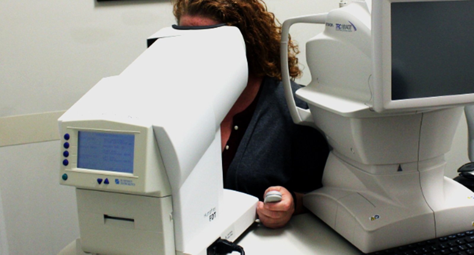
A visual field test is a diagnostic procedure used to assess the full extent of a person’s vision, both central and peripheral. It is commonly used in diagnosing and monitoring eye conditions such as glaucoma, macular degeneration, and neurological disorders that affect vision

Procedure: In a typical visual field test:

  • The patient is asked to focus on a central point (usually a light or a fixation point).
  • Stimuli, such as lights or patterns, will appear in different areas of the visual field.
  • The patient signals when they can see the stimuli, allowing the technician or doctor to map out the extent of vision loss.
  •  

Purpose: The visual field test helps in detecting areas of vision loss that might not be immediately noticeable, especially in conditions like glaucoma where peripheral vision is often the first to be affected.

Fundus Photo

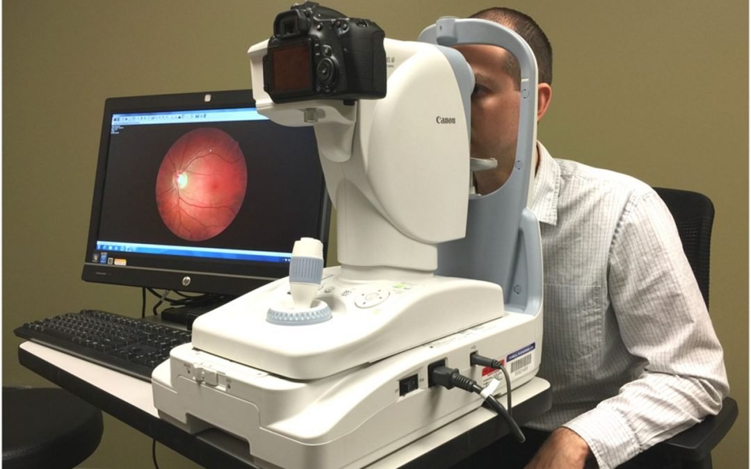
Fundus Photography is a diagnostic imaging technique used to capture detailed images of the fundus (the interior surface of the eye, including the retina, optic disc, macula, and blood vessels). This type of photography is crucial for assessing and monitoring a variety of eye conditions, particularly those related to the retina and optic nerve.

Applications in Eye Care

  • Screening: Fundus photography is widely used for screening at-risk populations, such as diabetics or elderly patients, for early signs of retinal disease.
  • Follow-up: It is often used in follow-up appointments to monitor the progression of retinal diseases, evaluate the success of treatments, and detect any new issues.
  • Pre-surgical Evaluation: For patients undergoing surgery, especially eye surgery, fundus images can provide essential information about the retina’s health before the procedure.

Yag Laser

YAG Laser (Yttrium-Aluminum-Garnet Laser) is a type of laser commonly used in ophthalmology for treating a variety of eye conditions. The YAG laser uses a crystal of yttrium-aluminum-garnet (YAG) as its medium, typically combined with a neodymium (Nd) dopant to produce laser light. This laser is non-invasive, highly focused, and used for both diagnostic and therapeutic purposes.

Advantages of YAG Laser

  • Non-invasive: YAG laser procedures are typically done with no incision, making them less risky and easier for the patient compared to surgical alternatives.
  • Quick and Effective: Most YAG laser treatments are quick and take only a few minutes to perform. Recovery times are generally short, and most patients can resume normal activities almost immediately.
  • Outpatient Procedure: YAG laser procedures are often done in an outpatient setting, meaning they do not require hospitalization.
  • Precise Treatment: YAG lasers allow for highly targeted treatment, minimizing damage to surrounding tissues.

OCT(Optical coherence tonography)

Optical Coherence Tomography (OCT) is a non-invasive imaging technique used to capture high-resolution, cross-sectional images of the eye, particularly the retina, optic nerve, and anterior segment. OCT is widely used in ophthalmology for diagnosing and managing a variety of eye conditions, especially those related to the retina and optic nerve.

Advantages of OCT

  • Non-invasive: Unlike other imaging techniques that may require injections or direct contact with the eye, OCT is completely non-invasive.
  • High Resolution: OCT provides extremely detailed images of retinal layers, optic nerve, and anterior segment, allowing for precise diagnosis and monitoring.
  • Quick and Comfortable: The procedure is quick, usually taking only a few minutes, and generally does not cause discomfort to the patient.
  • Real-Time Imaging: OCT provides real-time images that can be used immediately to evaluate conditions and guide treatment decisions.
  • 3D Imaging: Advanced OCT systems can provide 3D scans of the retina, offering a more comprehensive view and allowing for better monitoring of changes over time.

Autoref and keratometry

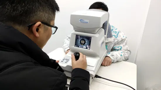
Both autorefraction (autoref) and keratometry are diagnostic tools commonly used in ophthalmology and optometry to assess the refractive error of the eye and the curvature of the cornea. These measurements are important for determining the best prescription for eyeglasses or contact lenses, and they help in evaluating conditions that affect vision, such as astigmatism, myopia, hyperopia, and more.

Uses

  • Astigmatism Diagnosis: Keratometry is essential for diagnosing astigmatism, a condition where the cornea or lens is irregularly shaped, causing blurred vision. It helps in determining the degree of astigmatism.
  • Refractive Surgery Evaluation: Before undergoing refractive surgeries like LASIK, keratometry is used to assess the corneal shape and curvature to determine if the patient is a suitable candidate for the procedure.
  • Monitoring Corneal Changes: It helps monitor corneal changes over time, particularly in patients with conditions like keratoconus (a progressive thinning and reshaping of the cornea).